November 2022
The annual workshop for teenagers who have been awarded an Arkwright Engineering Scholarship is always enjoyable. I couldn’t manage the weight of my Olympus BHMJ metallurgical microscope this time, so I took my Olympus SZ4045 stereomicroscope with my home-made shadowless illuminator and some metallic specimens.
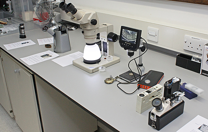
Assorted microscopes at the Arkwright Workshop
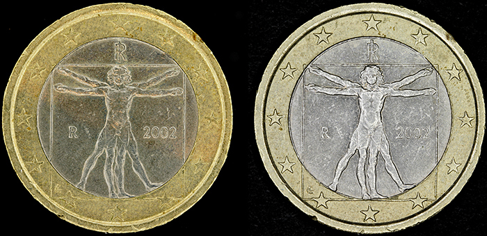
1 euro coin (left: shadowless illuminator, right: LED ring-light)
Metallurgical samples (a highly-polished metal surface appears black with shadowless illumination)
October 2022
I missed New Scientist Live and Quekex because we were in Thailand. We went to see one of Kai’s friends from school, and I was amazed to find that they had a large pond covered in duckweed and a lake big enough for large catfish. We were back in England in time for 2 outreach events, the National Honey Show at Sandown Park and Apple Day at Chinbrook Meadows. At the Honey Show, our stand backed on to GT Vision, so we had a good opportunity to examine their microscopes.
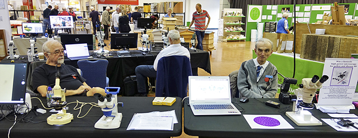
Quiet moment at the Honey Show
At Apple Day, we shared a stand with Thames21 again, and had lots of fun showing specimens from the River Quaggy to the local children.
Visiting families at Chinbrook Meadows
September 2022
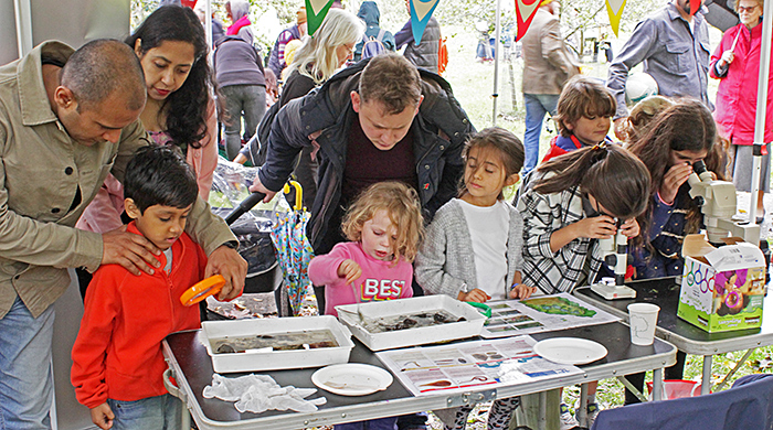
On Sunday 4th September, I joined Lisa and Nigel Ashby and Paul Smith at the Grove Park Carnival in Chinbrook Meadows in south-east London, where we shared the Thames21 area of the Friends of Chinbrook Meadows tent. We knew that there would not be an electricity supply, so we took an assortment of battery-powered microscopes and used them to show invertebrates collected from the River Quaggy, which runs through Chinbrook Meadows.
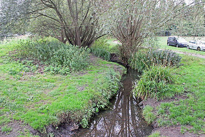
River Quaggy
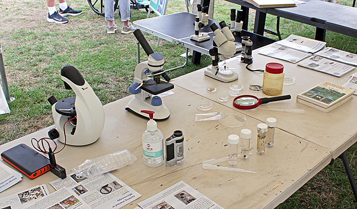
Microscopes and magnifiers
The specimens included mosquito larvae and pupae, freshwater shrimps (Gammarus), freshwater lice (Asellus), water fleas (Cladocera), copepods, a couple of bloodworms (larvae of non-biting midges, Chironomidae) and some small mayfly nymphs. The water fleas were pink, so they were probably Daphnia, which produce haemoglobin when the dissolved oxygen in the water is low. We used pipettes to transfer specimens to small Petri dishes for viewing under the stereomicroscopes or to microscope slides.
We had lots of visits from families, mostly with quite small children, and gave away lots of Fresnel magnifying lenses and leaflets on what to look at with microscopes, including the latest one on pond life.
On Saturday 10th September, I took some of the filters for my Olympus BH-2 and CH-2 microscopes to the workshop on filters and spectroscopy in the Natural History Museum. After seeing the narrow peaks produced by Pam Hamer’s dichroic filters with Paul Smith’s spectroscope, I expected a similar narrow peak at 550 nm from my IF 550 dichroic filter, but it gave a much broader peak, almost the same as my green absorption filter. The ND 25 dichroic neutral density filter also gave a disappointing result, clearly affecting some colours more than others.
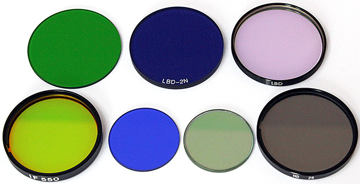
Olympus microscope filters
I was looking forward to the Wimbledon Common Open Day, usually an outreach event for Quekett members, but sadly it was cancelled because Queen Elizabeth died.
August 2022
I sorted out a few surplus filters, condensers and objective containers to sell at Microscopium in Letchworth on Saturday 13th, but I only sold one container because Mike Samworth and Phil Greaves had similar items at even lower prices than mine. It was not a wasted journey because Microscopium is always a good opportunity to meet up with old friends, and Phil was giving away card slide boxes so now I have enough boxes to arrange my slide collection better.
July 2022
On 1st July I had a sore throat, and the next day I felt quite ill and tested positive for Covid-19. I thought I had been careful about wearing a mask in shops and on buses and trains, and using hand gel, but obviously not careful enough. My symptoms persisted for a week, and I tested positive for 2 weeks.
June 2022
For the Home Counties Meeting at Pitsea on Saturday 11th June, I prepared a moss safari. I had been intrigued by the name of one of the Quekett’s followers on Twitter, Moss Safari, so I found the matching Moss Safari website produced by Andy Chandler-Grevatt. The website includes a procedure for collecting and soaking moss and then squeezing it and filtering the water before putting a drop on a slide. I collected moss from our garage roof 2 days before the meeting, soaked it, and took it to the meeting with my Olympus CH-2 microscope.

Moss Safari
Sadly almost everything I found was parts of moss. There were some very small creatures whizzing around, but they were so small that even with a 40× objective I couldn’t see what they were.
The next weekend was the Wimbledon Common Weekend of Nature (renamed from the BioBlitz). On Saturday 18th I went for the Butterflies & Dragonflies Walk and to take some photographs. On Sunday 19th, Barry Wendon, Neil Henry, Paul Smith and I set up our microscopes and cameras in the open-fronted garage next to the Information Centre to assist with identifying plants, insects and other organisms found during the organised walks. I took my Olympus SZ4045 stereomicroscope with a 144-LED ring-light, and used it to show various flowers, oak leaves with galls, a leaf with rust, lichens on an oak twig, and filamentous algae from the cattle trough.
Olympus SZ4045 stereomicroscope
One of the plants that we always see on the grassy plain is bird’s-foot trefoil (Lotus corniculatus), which gets its name from the similarity of the seed pods to the feet of a bird.
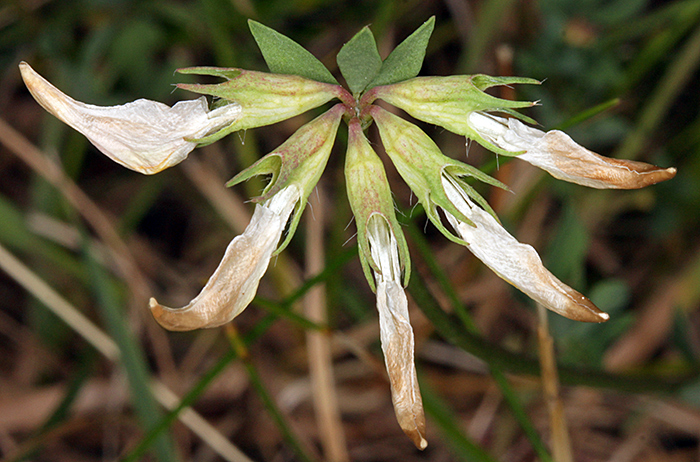
Seed pods of bird’s-foot trefoil
April 2022
This year is getting expensive. I had to replace my computer monitor in January, and now my 9-year old Dell XPS-8500 has died. The power light goes on and the fan starts, but the hard disk does not start, there are no beeps, no lights on the keyboard and only the BenQ logo appears on the monitor. And it would not boot from a USB drive. I think the BIOS or something else on the motherboard has failed, but the computer is too old to try to get it repaired. I was horrified at the price of a new Dell with a similar specification, so I chose an HP ProDesk 400 G7 with 16 GB RAM, 500 GB SSD (Windows and programs) and 1 TB hard disk (data). I can’t believe how quickly it starts, it is ready to sign in before I have folded the monitor dust cover. I had copies of most things on my laptop, so I was able to copy them to the new hard disk. I bought a cheap external hard disk enclosure and used it to transfer some other files from the Dell’s hard disk, which fortunately was still working, so I don’t think I have lost anything. I am now using an external hard disk for daily backups of my data and setup files.
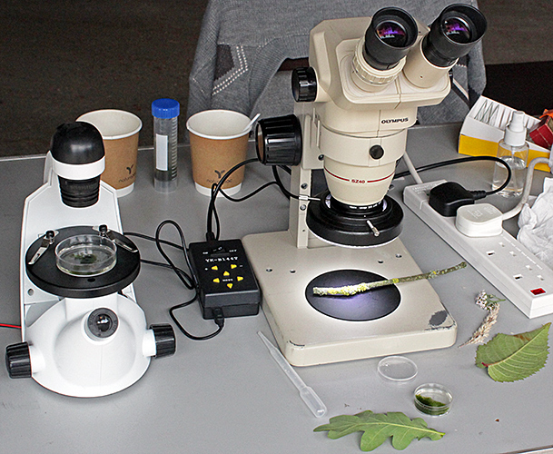
Dealing with the computer problems took a long time, so I was not able to prepare anything new for the workshop on contrast enhancement. I took the phase condenser and 4 phase objectives from my Olympus BHT microscope and mounted them on the Club’s Olympus BHC microscope so that I could demonstrate negative and positive phase contrast.
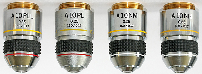
Olympus 10× PLL, PL, NM and NH phase contrast objectives
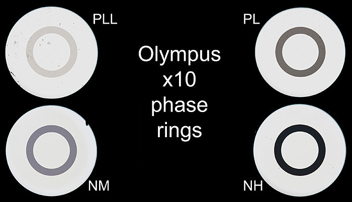
Phase rings in Olympus 10× objectives

Negative high (NH) and Positive low low (PLL) phase contrast
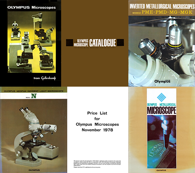
I also showed two ways to obtain dark-ground illumination for stereomicroscopes and macro photography that do not use optics.
One method uses an inverted LED ring-light with the aperture filled with black card. The other method uses an LED stage plate with a disk of black card obscuring most of it, leaving just a bright ring.
In both cases, the slide or specimen is placed on a piece of black foam board (with a central hole to let light through) resting on an inverted pudding basin from which the base has been cut out.
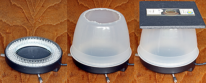
Dark-ground illuminator, showing (left) black disc, (centre) inverted bowl with base removed, and (right) the complete illuminator
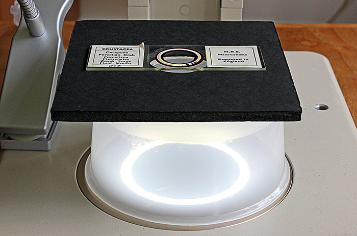
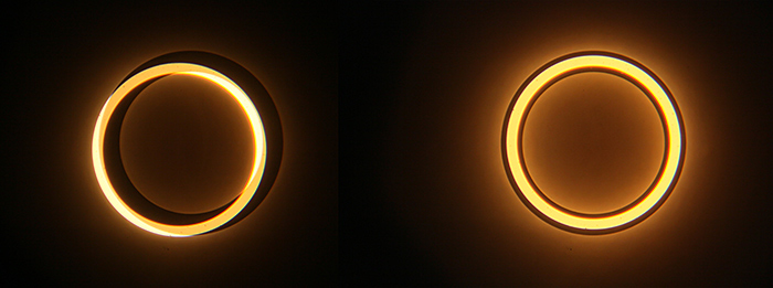
Dark-ground illuminator using an LED stage plate
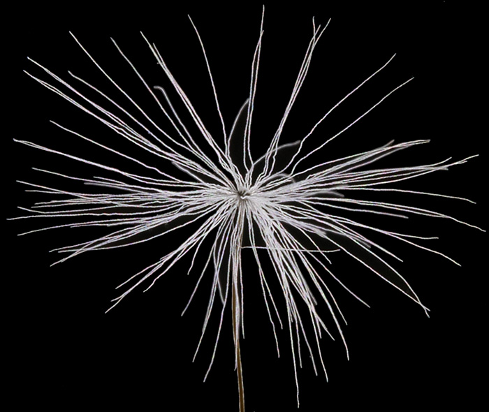
Seed of dandelion (Taraxacum officinale)
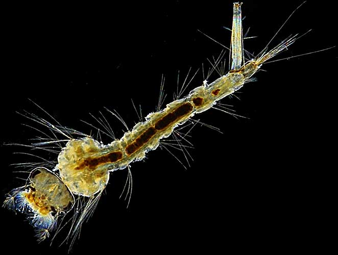
Larva of a mosquito (Culex pipiens L.) on a slide by T. Gerrard & Co.
It was good to meet up with old friends at the Reading Convention. It felt strange to be there without a camera, because Peter Wyn-Jones (the new webmaster) was there taking photos for his report, but it gave me more time to talk to people and browse all the items for sale. I didn’t manage to prepare an exhibit, but I took a few items to sell and I came home with a wooden slide box and more money than I started.
March 2022
On 16th March, dust from the Sahara Desert was blown over southern England and it rained so that the dust was washed out of the air and deposited everywhere. The deposits had dried by the next day so I collected some from the top of one of our recycling bins. Now I have to try to find time to at it under one of my microscopes.
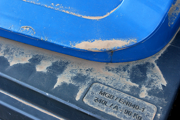
I have finally managed to catch up with scanning documents for the Olympus microscope documentation section of my website. Some of these came from Mike Samworth at Penkridge in September, where he was disposing of items from the library of the Postal Microscopical Society. Others came from Hampshire Micro and were donated by Dave Routledge.
Olympus catalogues and brochures
For the gossip following the AGM (where Peter Wyn-Jones took over as webmaster), I prepared some notes on phase telescopes, with photographs of the phase ring in the back of an objective and the phase annulus in the condenser, before and after alignment with the screws on my Olympus BH2-PC condenser.
Left: Before alignment Right: After alignment
I had to take the photographs through the phase telescope, so it was not possible to use a photo eyepiece; I used my Canon EOS 5D Mark II with an old Olympus Zuiko 50mm f/1.8 standard lens, supported on a copy stand.
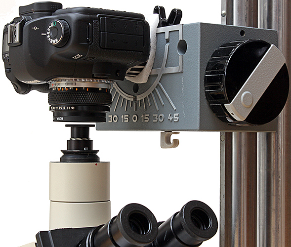
Afocal arrangement for photographing phase rings
I first needed a phase telescope when I bought my Olympus CK2 inverted microscope on eBay in 2010, and the seller very kindly gave me a Watson one. I soon decided that I wanted phase contrast on my BH-2 as well, so I bought a BH2-PC condenser and started collecting objectives. I wanted to have a phase telescope for the BH-2, and real Olympus ones were expensive, so I bought another Watson because it was cheap. As time passed, I found an Olympus CT-4 and then a CT-5 at reasonable prices, so I will probably sell the Watson ones. I don’t understand why the Watson ones are so big.
Olympus CT-4, Watson and Olympus CT-5 phase telescopes
February 2022
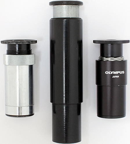
For the 9 years that I have been webmaster and writing meeting reports for the website, I have never had a good way of photographing slides at meetings. Several Quekett members use an afocal arrangement with a Leitz Periplan 10× eyepiece that has a 28mm thread (originally for a rubber eyecup) that makes it easy to attach to the filter thread of a camera lens, and I found one at Microscopium a few years ago. Last month I finally ordered a step-down ring from a Chinese seller on eBay so that I could start experimenting, and I showed the results on my laptop at the Home Counties Meeting at Brookwood.
Alan Wood’s exhibit
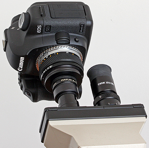
Afocal coupling of EOS digital SLR, Zuiko 50mm standard lens and Periplan 10× eyepiece
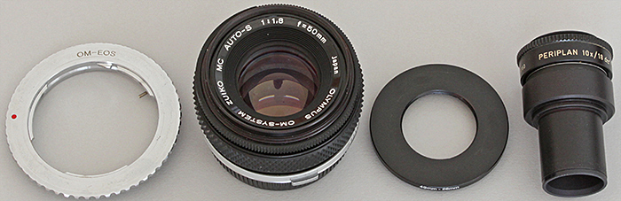
OM-EOS adapter, Zuiko 50mm lens, 49–28mm step-down ring, Periplan 10× eyepiece
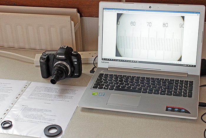
I prepared a PowerPoint presentation to show images of a stage micrometer and a section of a swan mussel that I used to compare an afocal arrangement using a Periplan 519750 with a conventional setup using an NFK 2.5× photo eyepiece:
Click the arrows to move through the slides. Click the symbol at bottom right for a larger version.
January 2022
Another month with no time for microscopy. I am still working on the revision of ISO 1750 Pesticides and other agrochemicals – Common names, not helped by an unscheduled trip to Thailand to sort out some problems. I did manage to almost finish work on the IUPAC systematic names, and to update hundreds of SVG images of structural formulae to indicate that they are racemates. I was looking forward to the “My latest microscopical acquisition” gossip meeting, but we were in Thailand where the Zoom meeting started at 1:00 a.m., too late for me. However, I hope to find time to watch the recording on the Quekett website. I hope to have some time for microscopy in February.
When I started working on the Quekett website back in 2012 I realised that I would be editing lots of photographs, so I treated myself to a 24″ Dell U2410 Ultrasharp with a 1920×1200 IPS screen that came colour calibrated from the factory. I prefer the 1200 height to the more common 1080. It has worked perfectly until this month when the screen went black as usual during the Windows Update but stayed that way. I tried the Dell monitor with another computer and it did not show an image, so I decided it was time for a new monitor. I looked at the Dell Ultrasharp range, but the 24″ models no longer come calibrated. Google found the SW240 from BenQ, a brand I had not previously considered, with an 1920×1200 IPS screen and a calibration certificate, and I bought one. The display is just as good as the Dell, and it runs so cool that I can cover it as soon as I turn it off; the Dell took a long time to cool down.
Commenting on this blog
If you would like to comment on anything in this blog, please send me a message.