December 2014
I have had hardly any time for microscopy this month, but here is something with a Christmas theme:
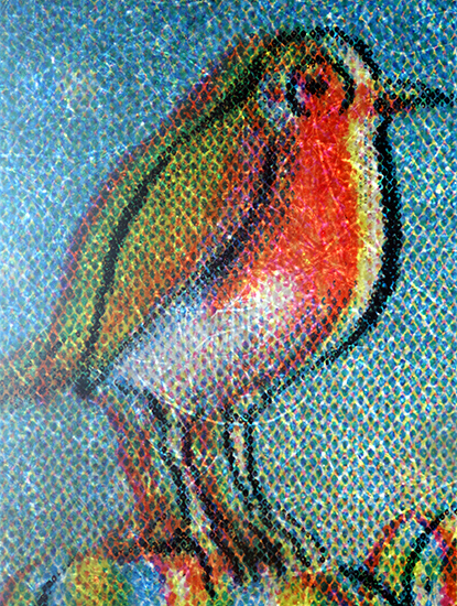
Robin on 2013 UK 1st class Christmas stamp
Olympus SPlanFL 2× objective and NFK 2.5× photo eyepiece, stack of 8 images in Zerene Stacker
November 2014
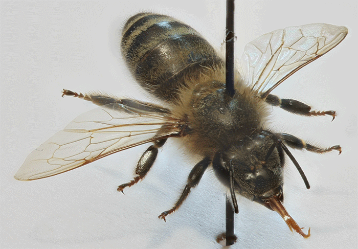
Norman Chapman, Pam Hamer and I manned the Quekett stand on the final day of the National Honey Show on Saturday 1st November. We had specimens of honeybees, Varroa destructor Anderson & Trueman, and the small hive beetle (Aethina tumida Murray) and slides of pollen, and after the meeting I took photographs of them to use in the meeting report.
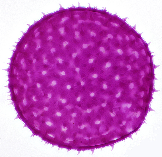
Stained single grain of pollen of mallow (Malva sp.) from an NBS slide, diameter 150 µm
Olympus SPlan 40× objective, NFK 2.5× photo eyepiece, stack of 12 images in Zerene Stacker
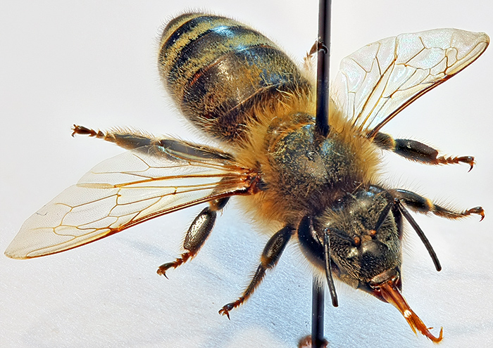
Pinned specimen of a honeybee (Apis mellifera L.)
Canon EOS 40D with 60 mm EF-S macro lens at f/4.5, stack of 25 images in Zerene Stacker, focus steps using EOS Utility
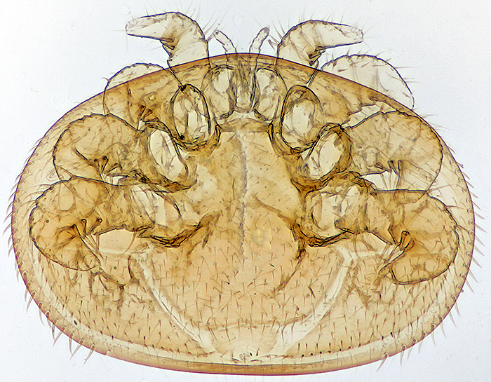
Cleared female Varroa destructor Anderson & Trueman, mite is 1.65 mm wide, slide by L & P Bircham
Olympus SPlan 4× objective, NFK 3.3× photo eyepiece, stack of 22 images in Zerene Stacker
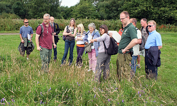
Tim Newton, Dennis Fullwood and I manned the Quekett stand at the annual exhibition of the British Entomological and Natural History Society in Conway Hall in London on Saturday 8th November, the first time that the Club has had a stand at this event. We wanted to show entomologists how microscopes can make their lives easier, so I had prepared some insects on a setting board using my Olympus SZ4045 stereomicroscope. This brought back memories; I was a teenager collecting butterflies the last time I set any insects, and I hadn’t even heard of stereomicroscopes then.
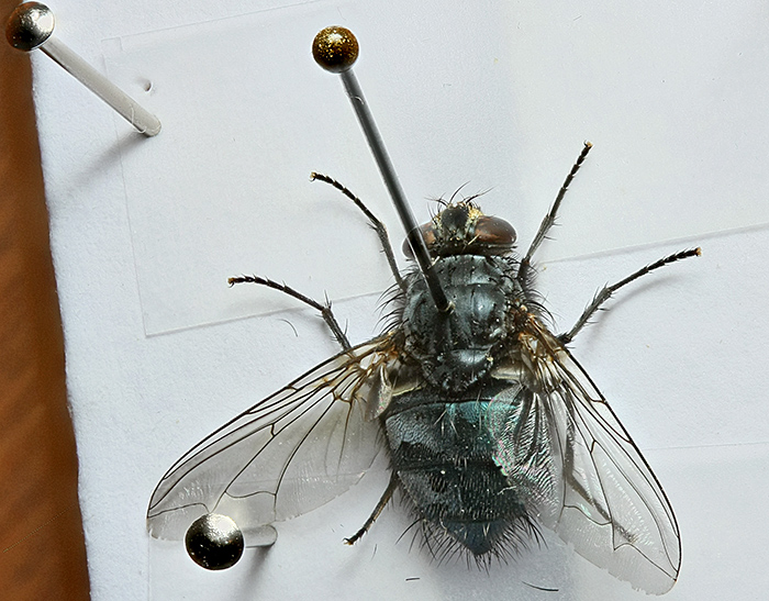
Fly arranged on a setting board
Canon EOS 40D with 60 mm EF-S macro lens at f/4.5, stack of 37 images in Zerene Stacker, focus steps using EOS Utility
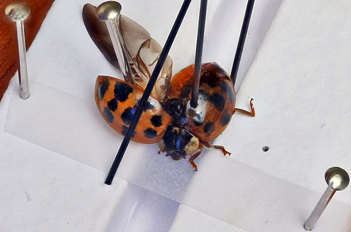
Harlequin ladybird (Harmonia axyridis (Pallas)) with one wing extended, on a setting board
Canon EOS 40D with 60 mm EF-S macro lens at f/4.5, stack of 30 images in Zerene Stacker, focus steps using EOS Utility
Alan Cooper’s presentation at the joint workshop with the Stereoscopic Society on Saturday 15th November reminded me that Zerene Stacker can extract synthetic stereo images from a stack and prompted me to have a go. Here is a rocking animated GIF of a pinned honeybee:
Rocking animation of a honeybee (Apis mellifera L.), images generated by Zerene Stacker and combined into an animated GIF in Photoshop Elements
Alan Cooper recommended StereoPhoto Maker for producing stereoscopic images, so I exported 2 images from Zerene Stacker as a stereo pair and then used StereoPhoto Maker to produce images for free viewing and for red/cyan 3-D anaglyph glasses. I didn’t even need to read the StereoPhoto Maker Help.
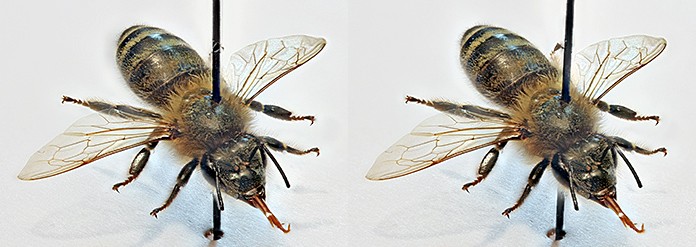
Stereoscopic pair of images of a honeybee for free viewing
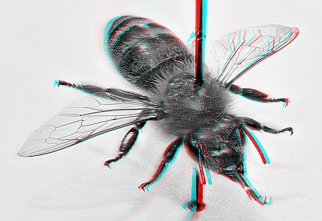
Stereoscopic image of a honeybee for viewing with red/cyan anaglyph glasses
October 2014
The photo of a common spangle gall that I took in September was chosen to be displayed at the annual exhibition on Saturday 4th October, and I got a Barnard certificate for it. For my exhibit, I showed how defects in a crystal can be made very obvious using dark-ground illumination under a stereomicroscope, and how plastic films from greetings cards and food packaging can be used as waveplates to change the colours of crystals under crossed polarisers.
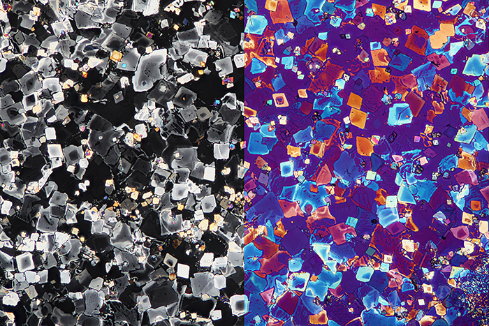
Potassium chlorate crystals under crossed polarisers (left) and with a plastic film as a waveplate (right)
Olympus SPlan 4× objective, NFK 2.5× photo eyepiece, stack of 10 images in Zerene Stacker
Mark Papp and I manned the Quekett stand at the Antique Scientific Instrument Fair on Sunday 26th October. There were some fascinating microscopes for sale, but at very high prices. However, it was great to be able to admire the workmanship in the old brass microscopes, and the ingenious ways that some of them had been designed to fit into tiny wooden boxes.
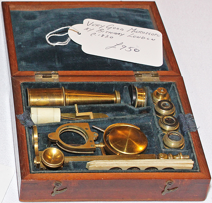
Dismantled Bithray brass microscope in its fitted wooden box
My attempts at DIY don’t often turn out very well, but here is one that did. I wanted a 5-way nosepiece to put on my Olympus CH-2 without borrowing one from my BH-2, and I obtained one from a wrecked BHTU, but its rubber grip was missing. It has taken me some time to find a suitable material, but now I have found a sheet of corrugated black rubber at the 4D Model Shop, not far from the Tower of London. It is 3 mm thick, which is just right. It was easy to cut a thin strip using a rotary trimmer, and fairly easy to stick it in place with UHU adhesive. I used a rubber band to hold it in place while the adhesive set.
Replacement rubber grip on replacement 5-way nosepiece on Olympus CH-2 microscope
Canon EOS 40D with 60 mm EF-S macro lens at f/4.5, stack of 16 images in Zerene Stacker; I used EOS Utility to make the focus steps
September 2014
The annual exhibition is approaching fast, so I must get my photos to David Linstead by 20th September. I will submit the nettle hairs that I shot in August, and I have plans for some others.
While I was in Thailand at the beginning of the month, I saw an item on the television news about processing blanket weed (filamentous algae) into small pellets and then using it to colour (pale green) and flavour ice cream in Chiang Mai. I have heard of Chlorella being hailed as a superfood, but not Spirogyra!
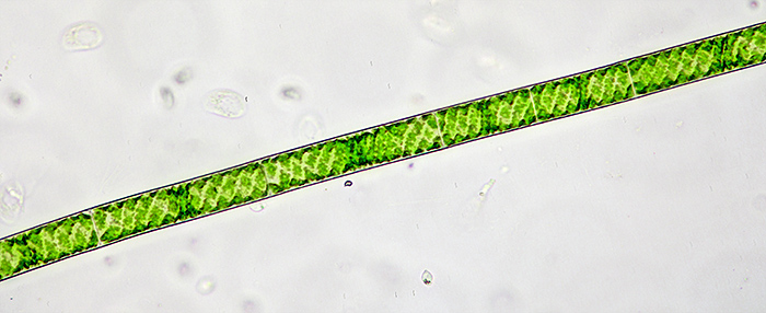
Single filament of Spirogyra sp. from Queensmere, showing the green chloroplasts arranged in a spiral
Olympus SPlan 20× objective, NFK 2.5× photo eyepiece, stack of 17 images in Zerene Stacker
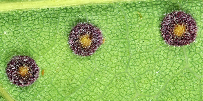
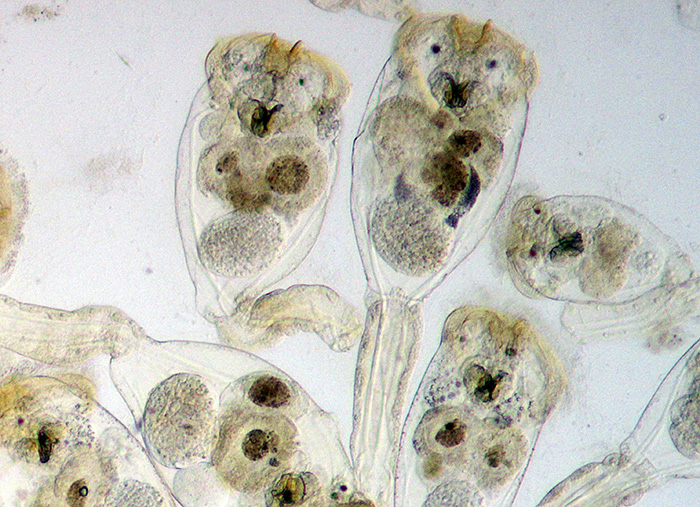
Sunday 14th September was Stables Open Day on Wimbledon Common, and Dennis Fullwood, Paul Smith, Barry Wendon and I were in the Information Centre with our microscopes and cameras. This was the busiest Quekett event I have been to, and I reckon we had over 200 visitors, far more than in previous years. Waterfleas and other pond life from Queensmere normally get the most attention, but this year spangle galls (caused by Cynipid wasps) on oak leaves were the star attraction.
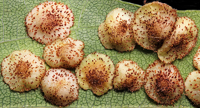
Common spangle galls, caused by Neuroterus quercusbaccarum (L.)
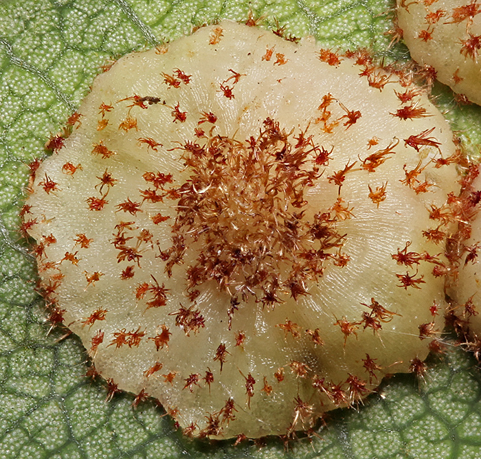
Common spangle gall
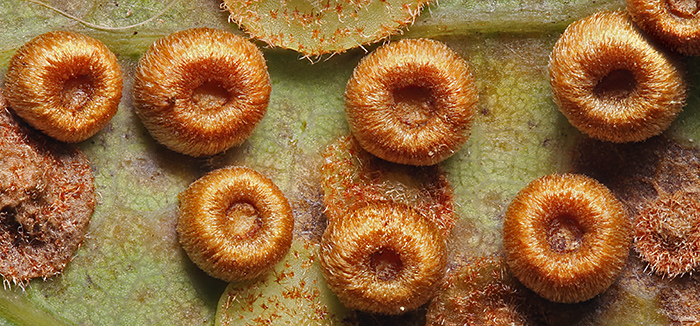
Silk button spangle galls, caused by Neuroterus numismalis Geoffroy in Fourcroy
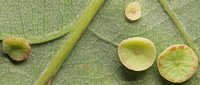
Smooth spangle galls, caused by Neuroterus albipes Schenck
I have sent a larger version of the single common spangle gall off to David Linstead. All 4 photos were taken with my Canon EOS 600D and 60 mm EF-S macro lens, with an additional +8 dioptre close-up lens for the single gall; I used EOS Utility to make the focus steps. They are all stacks of about 20 images in Zerene Stacker.
August 2014
On Saturday 2nd August, I went to the Rock Gem ‘n’ Bead Show at Kempton Park and bought a couple of specimens for my exhibit at Quekex (Saturday 4th October, with crystals as the main topic). As usual, Dennis Fullwood was there with the Kent Geologists’ Group and half a dozen microscopes, encouraging visitors old and young to try their hand at mounting microfossils.
On Saturday 9th August, I didn’t take a trinocular microscope on the Quekett excursion to Warnham Local Nature Reserve because it is a 25-minute walk from Horsham station, and then wished that I had made the effort because there were so many rotifers in the samples from the dipping ponds. In the afternoon I spotted a common lizard sunning itself on a fence, and I managed to get close enough to photograph it.
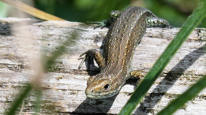
Common lizard (Zootoca vivipara (Lichtenstein)) at Warnham Local Nature Reserve
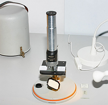
The next day, Sunday 10th August, as part of the Club’s outreach programme Graham Matthews and I were back at the Reserve for their I-Spy Nature Day aimed at local families. I took 2 microscopes that are suitable for children, a new Chinese stereo with fixed 20× magnification and built-in LED illumination for under £40 from eBay seller gt-vision, and my old Meopta portable compound (with Ikea Jansjö LED lamp).
Stereo and compound microscopes suitable for children
With pond life left over from the previous day’s excursion and specimens that people brought to us, we had plenty of material to educate and entertain a good crowd of children and parents, including stinging nettles.
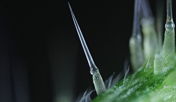
Hairs on stem of stinging nettle (Urtica dioica L.)
Olympus SPlan 4× objective, NFK 2.5× photo eyepiece, stack of 24 images in Zerene Stacker
July 2014
I am hopeless at public speaking, but I managed to stumble through the introduction to the Quekett gossip on “My Latest Microscopical Acquisition” on Tuesday 8th July, with a PowerPoint presentation listing the international eBay categories that I have found useful for microscopes, and translations of useful phrases such as “Time: newly listed” and “Save search”.
On Saturday 13th July, I took my Olympus SZ4045 stereomicroscope to the first Wimbledon Common BioBlitz. Paul Smith, Dennis Fullwood and Barry Wendon were there too, as part of the Club’s outreach programme. At the next BioBlitz on the Common, there are plans to include organised pond-dipping in Queensmere, encouraging visitors to bring their finds to the Centre to examine them with our microscopes.
Guided walk on Wimbledon Common
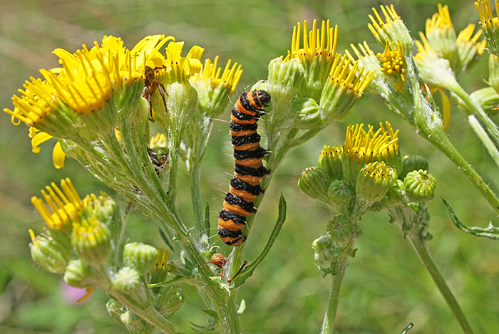
Caterpillar of cinnabar moth (Tyria jacobaeae (L.)) on ragwort (Jacobaea vulgaris Gaertn.) on Wimbledon Common
Canon EOS 40D, EF-S 18-55 mm zoom at 37 mm, f/11, 1/500 sec, ISO 500, hand-held
Galls on leaf of oak (Quercus sp.) from Wimbledon Common
To complete my trinocular compound microscope that is light enough to carry on public transport, I bought a refurbished Canon EOS 600D camera from eBay seller canon_uk1. I chose this camera model because it is the lightest one with an articulated LED screen that can face forwards when the camera is pointing down. The screen isn’t level in the photograph below because Canon have restricted the rotation of the screen to 175°; I don’t understand why.
Canon EOS 600D digital SLR camera on Olympus CH-2 trinocular microscope
On Saturday 26th July, I took my Olympus CH-2 and EOS 600D to Brookwood Memorial Hall for the Quekett’s excursion to the Basingstoke Canal, the first time I have used them away from home. Everything worked, but it is definitely not as easy as the experts like Graham Matthews and Tony Pattinson make it look.
Conochilus sp., a colonial rotifer, squashed by the coverslip, from the Basingstoke Canal
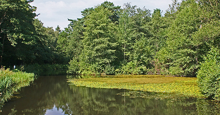
Basingstoke Canal, near Pirbright Bridge
June 2014
We spent 3 weeks in Thailand, but as usual family problems took nearly all of our time. We did manage to get to the seaside for a few hours and I collected sand from the beach at Cha-am on the west coast of the Gulf of Thailand for Paul Smith. The sand looked quite pale so I was hoping for some forams, but the only biological material seems to be bits of shell.
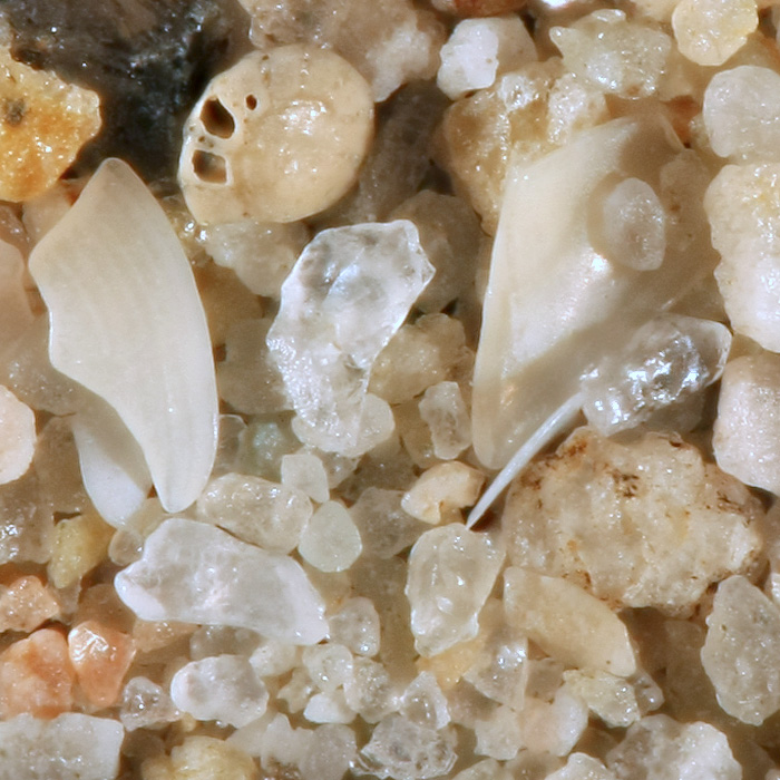
Sand from the beach at Cha-am in Thailand (the circular shell at the top is about 1 mm across)
At the gossip meeting “NBS – Products and Reminiscences” on Tuesday 10th June, James Rider and Kit Brownlee each showed an NBS Polarspex but they were not photographed. I couldn’t attend the meeting, so I photographed my Polarspex for inclusion in the meeting report.
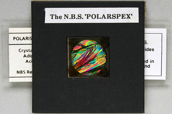
NBS Polarspex with a slide of adipic acid crystals
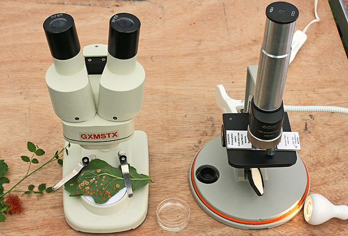
Saturday 21st June was the Quekett Microscopical Club’s Young Scientists’ Day based in the Angela Marmont Centre in the Natural History Museum. I took an assortment of specimens and my 2 smallest microscopes, an old Meopta portable compound (with Ikea Jansjö LED lamp) and a new Chinese stereo with fixed 20× magnification and built-in LED illumination for under £40 from eBay seller gt-vision.
 |
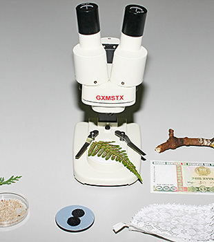 |
| Meopta portable microscope | GXM STX 20× stereomicroscope |
Chasing pondlife from the Museum’s Wildlife Garden around a 55 mm Petri dish under a stereomicroscope reminded me to look for some smaller dishes on eBay. I found only one seller with smaller dishes, and ordered a pack of ten 30 mm ones from Hong Kong. They came quickly, but they are not as small as claimed; the base is 35 mm and the top 40 mm. The field of view of my Olympus SZ4045 stereomicroscope at its lowest magnification (6.7×) is 35 mm, so I can just see a whole dish at once.
May 2014
Here is something you don’t see very often – a photograph of me – because I am normally taking the photographs. This was taken in the Information Centre on Wimbledon Common during the Club’s excursion on Saturday 17th May.
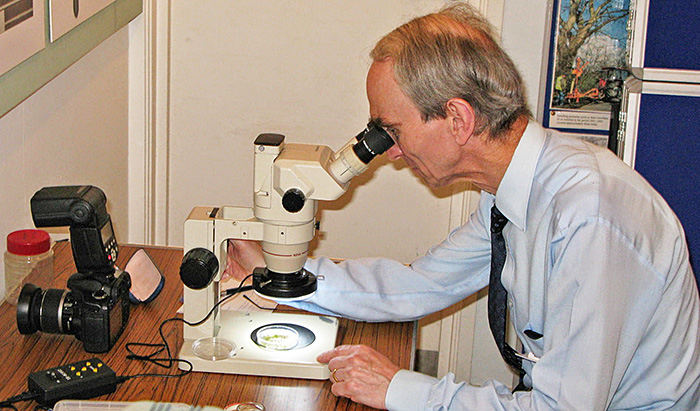
Alan Wood with his Olympus SZ4045 trinocular stereomicroscope [by Graham Matthews]
My Olympus BH-2 is too heavy for me to take it on Quekett excursions by public transport (I can’t drive), so I have been looking for something lighter. I took a chance on a CH-2 stand that someone had found in their garden shed. Cleaning out the dead leaves, insects and spiders was easy, and the paintwork cleaned up nicely with a paste of sodium bicarbonate, but the coarse focus would not move. I found that a previous owner had dissolved out all of the lubricant and turned the tension all the way up. Several turns of the tension control got the focus moving enough to start introducing some lubricant from OSIM Optik Shop, and I soon had a functioning stand. At the 2013 Reading Convention, Tony Pattinson had convinced me that it was easy to remove the 4-way nosepiece and replace it with a 5-way one from a BH-2 after removing the dovetail, and he was right; the screws were very tight but I managed to undo them with the proper JIS screwdriver. However, I found that not all nosepieces can be fitted to a CH-2; you need to find one that has 2 empty screw holes, one on each side of the removable dovetail, as in the left picture below.
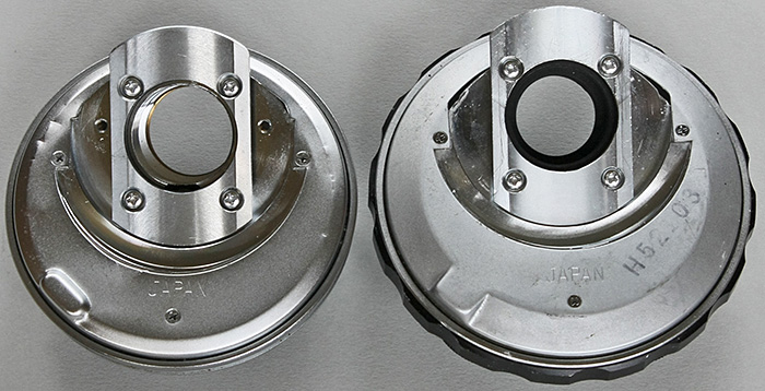
Nosepieces from an Olympus BH-2; only the one on the left has the extra holes needed for fitting to a CH-2
I still had the set of Olympus A objectives that came with my BH-2 (now upgraded to S Plans) and some spare WHK eyepieces, so I now have a complete and working CH-2 with 5 objectives. Now I need a lighter camera to go with it.
March 2014
I have been working on a demonstration of the 4 different types of phase contrast that Olympus provided for the BH-2 and other microscopes of that generation; PLL (positive low low), PL (positive low), NM (negative medium) and NH (negative high). I got the photographs ready just in time for the 25th meeting of the South Thames Discussion Group at Cobham on Saturday 1st March, and used the same setup again at the 2014 Reading Convention on Saturday 8th March.
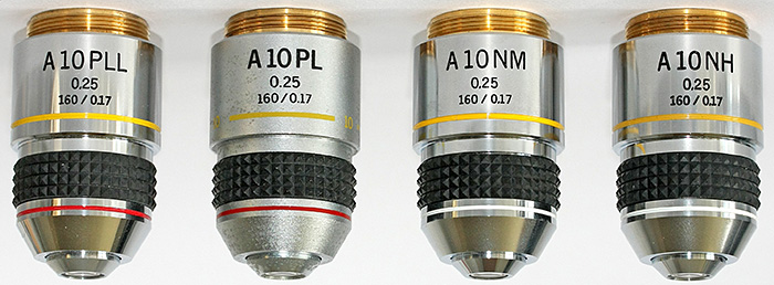
Olympus 10× PLL, PL, NM and NH phase contrast objectives
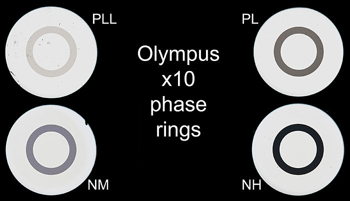
Phase rings in Olympus 10× objectives

Negative high (NH) and Positive low low (PLL) phase contrast
I can’t carry my BH-2 on public transport, so I took my Olympus CK2 inverted microscope instead, with a monocular head and plain stage to keep the weight down.
January 2014
Martin Hinchcliffe sold all of his beautifully-made brass specimen holders at Microscopium in October before I had a chance to buy one, but he has made a new batch and I have now bought one.
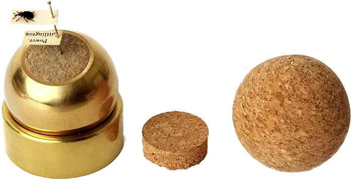
Martin Hinchcliffe’s specimen holder
It comes with one 30 mm cork ball, one 30 mm brass ball with a cork insert, and 2 spare inserts. The weight of the brass ball and the friction of the cork one keep specimens at the angle you want under a stereomicroscope or a camera.
Commenting on this blog
If you would like to comment on anything in this blog, please send me a message.